animal models /
pig models
/

pig models

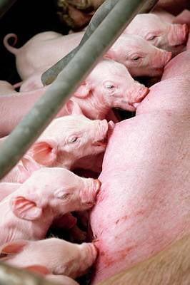
The domestic pig has served as an important agricultural resource and as an excellent large biomedical model for translational research due to its anatomical size and similar physiology to humans.  In contrast to mouse model generation, genetic modifications in pigs have been hampered because of the lack of characterized embryonic stem cells (ESCs), the extremely low efficiency of homologous recombination (HR) and time-consuming breeding programs necessary to produce biallelic genetically modified animals. The derivation of stable lines of embryonic stem cells from swine with germline transmission potential has been difficult to achieve in many labs. This technical obstacle precludes an ES-cell mediated route for animal model generation in the domestic pig. A recent noteworthy report originating from Pentao Liu’s laboratory has described the generation of expanded potential stem cells (EPSCs) from individual pig blastomeres that have analogous signaling requirements to human EPSCs and can successfully differentiate into primordial germ-like cells (PGCLCs). While derivation of these expanded potential stem cells represents an exciting milestone, at the time of writing, no live born chimeric EPSC piglets have been produced with demonstrated germline transmission (See Gao et al, 2019¹,²). Alternative approaches to generate porcine animal models such as genetic reprogramming with Yamanaka factors OCT4 (POU5F1), SOX2, KLF4 and c-MYC to induce nuclear reprogramming to naïve pluripotency have generally failed to produce stable, karotypically normal pig iPSC lines that are independent of the transgenic reprogramming factors. As such, porcine fibroblasts have been used generally as the target cells for gene editing because they are amenable to mammalian cloning by somatic cell nuclear transfer (SCNT) to generate pig biomedical models. In spite of the epigenetic barriers that impede reprogramming (Matoba & Zhang 2018³ ), mammalian cloning by SCNT has become the established gold-standard biotechnology for production of large animal models including cows, goats, pigs and sheep (Estrada et al 2008⁴).

For decades, targeted genetic modification in swine and other large biomedical models had been notoriously tricky because homology-directed repair (HDR) occurs at a such a low frequency (often 1 in 10,000) in fibroblasts in comparison to gene targeting frequencies in traditional mouse ES cells (~1 in 100). With the advent of site-specific gene editing tools, chiefly CRISPR-Cas, a DNA double-stranded break (DSB) is intentionally introduced at the target site to stimulate homologous recombination (HR) with a frequency 50-1000-fold higher at the intended locus.  This increased HR activity results in a high rate of modification and, thus, reduces the number of fibroblast colonies to be screened by long-range PCR for the targeted allele.  The CRISPR-Cas technological platform accelerates the potential for seamless modifications by gene knockout and knockin.  Indeed, recent genome engineering efforts in the domestic pig have inactivated multiple immune targets and all 62 copies of porcine endogenous retroviruses (PERVs) with the goal that swine organs such as the heart or kidneys can be tolerated without humoral rejection and transmission of pathogens to humans (Fischer et al 2016⁵ & Niu et al 2017⁶). Multiple companies including NovoHelix are advancing porcine xenotransplantation efforts to alleviate the shortage of donor organs for transplantation into humans.  At NovoHelix, we have optimized gene editing techniques to address the critical need for large animal models across a variety of disciplines in biomedicine and translational research.  Please contact us through our online quoting system for project quotes and consultation. 
model generation
support services
technology
references
forms
quote request
model generation

Service

Catalog Nr

Service Description

Timeline

Deliverables

Pricing

Custom Genetic Modification - Pig Biomedical Model
GESS001
NovoHelix scientists provide regular updates for porcine model generation milestones including isogenic targeting vector construction, establishment of primary cell lines from elite animals for somatic cell nuclear transfer (SCNT) and genome engineering, validation and expansion of single-cell gene-edited clones, transfer of SCNT embryos to recipient surrogates, pregnancies established and gestational outcomes, and delivery of the final gene-edited founder animals.
9- 24 months
 2 founders with the desired genetic modification
support services

Service

Catalog Nr

Service Description

Timeline

Deliverables

Pricing

  • Gene editing activity testing
  • Format - cell-based transfection
  • Assay - T7 endonuclease I/Cel-II/Surveyor
 GESS004
NovoHelix offers a gene editing service to help clients test their CRISPR tools including guide RNAs, high-performance mutant Cas proteins and base editors in plasmid DNA or RNP formats.  A representative cell line will be transfected in triplicate and results will be generated by the mismatch-nucleases T7 Endo I or Cel-II as adopted from a protocol originally developed by Keith Joung's lab. While our cell-based assay is the gold standard for guide RNA activity, guide RNAs can be tested in an alternative in vitro cutting assay should the client prefer.  However, caution must be used in interpreting these in vitro results, as NovoHelix has broadly found that Cas9 RNP in vitro cutting of PCR amplicons as a surrogate assay for gRNA activity vastly overrepresents Cas RNP in vivo activity levels both in cellular and microinjection contexts. Hence in vitro mismatch nuclease assays often do not correlate to gene editing in vivo outcomes. In short, the cell-based gRNA testing is strongly suggested before proceeding to animal model generation.

 7-10 days

 Gene editing activity of up to 6 guide RNAs

  • Gene editing activity testing
  • Format - cell-based transfection
  • Assay - genetic reporter & flow cytometry
GESS005

 NovoHelix offers a gene editing service to help clients test their CRISPR tools including guide RNAs, high-performance mutant Cas proteins and base editors in plasmid DNA or RNP formats.  A representative cell line will be transfected in triplicate and results will be generated by a fluorescent reporter assay and flow cytometry.

 7-10 days

 Gene editing activity of up to 6 guide RNAs

 Genotyping Assay Development - conditional knockouts
 GESS006
NovoHelix offers a knockout genotyping service to help clients develop robust genotyping protocols for screening animals after breeding and expansion of indidvidual mutant/cKO founder lines.

 2-4 days

 genotyping protocol

 development of guide RNAs - guide RNA cloning and design in a mammalian (U6, H1) expression vector or T7 in vitro transcription (T7- IVT) vector (DNA format)
GESS007

 Service is for design and cloning of up to 6 guide RNAs for 1 target locust.

 7-10 days

 6 guide RNAs in plasmid vectors

 development of guide RNAs - guide RNA cloning and design with T7 in vitro transcription (T7- IVT) for use in RNP format (RNA format)
GESS008
 Service is for design and in vitro transcription of up to 6 guide RNAs for 1 target locust.

 7-10 days

 6 guide RNAs ~ 500 ng/ul --- RNA format

 client-provided guide RNAs - preparation for microinjection (DNA/RNA format)
GESS009
Service is for purification and for preparation of gRNAs for zygotic microinjection or slide-electroporation. Client should provide validated gRNA synthesis data such as small RNA electrophoresis results from Agilent's Bioanalyzer prior to microinjection and a minimum 200 ng/ul gRNA concentration. Purification is required to prevent the microinjection glass needle from clogging, to prevent embryo lysis and extensive delays in refitting and resetting microinjection setup.
 client-provided dsDNA donor vector - preparation for microinjection 
GESS010
Purification of supercoiled plasmid by a proprietary anion exchange (AEX) chromatography, validation by restriction digestion and analysis by gel electrophoresis. Endotoxin levels are generally very low (0.05  – 0.5 ng LPS/µg) via our proprietary extraction method and are adequate for sensitive applications such as zygotic microinjection of mammalian embryos. The plasmid is predominantly in its supercoiled topology and free of RNA and protein contamination such as RNases and proteases.  Drop dialysis in nucleofection/electroporation/microinjection buffer is included in the purification service. Validation by additional restriction digestions (greater than 3) is available with commensurate fee schedule.  Purification is compulsory to prevent the microinjection glass needle from clogging, to prevent embryo lysis and extensive delays in refitting and resetting the microinjection setup.
 dsDNA donor vector construction
GESS011
Isogenic cKO dsDNA donor vector construction and Sanger-sequence verified with up to 5-kb floxed insertion.  NovoHelix will provide an in silico map and provide restriction fingerprinting for plasmid structure via 3 digests. DNA is purified by a propriety AEX (anion exchange) chromatography and suitable for microinjection into mouse zygotes or nucleofection/electroporation.  The DNA is dialyzed on a membrane support in microinjection or electroporation buffer and then spun to remove particulate matter to obviate microinjection needle clogging.
 dsDNA donor vector construction - complex
GESS012
 Isogenic cKO dsDNA donor vector construction and Sanger-sequence verified with up to 20-kb floxed insertion.  NovoHelix will provide an in silico map and provide restriction fingerprinting for plasmid structure via 3 digests. DNA is purified by a propriety AEX (anion exchange) chromatography and suitable for microinjection into mouse zygotes or nucleofection/electroporation.  The DNA is dialyzed on a membrane support in microinjection or electroporation buffer and then spun to remove particulate matter to obviate microinjection needle clogging.
technology
references
1: Gao X, Nowak-Imialek M, Chen X, Chen D, Herrmann D, Ruan D, Chen ACH, Eckersley-Maslin MA, Ahmad S, Lee YL, Kobayashi T, Ryan D, Zhong J, Zhu J, Wu J, Lan G, Petkov S, Yang J, Antunes L, Campos LS, Fu B, Wang S, Yong Y, Wang X, Xue SG, Ge L, Liu Z, Huang Y, Nie T, Li P, Wu D, Pei D, Zhang Y, Lu L, Yang F, Kimber SJ, Reik W, Zou X, Shang Z, Lai L, Surani A, Tam PPL, Ahmed A, Yeung WSB, Teichmann SA, Niemann H, Liu P. Establishment of porcine and human expanded potential stem cells. Nat Cell Biol. 2019 Jun;21(6):687-699. doi:10.1038/s41556-019-0333-2. Epub 2019 Jun 3. PubMed PMID: 31160711.

2: Li H, Zhao C, Xu J, Xu Y, Cheng C, Liu Y, Wang T, Du Y, Xie L, Zhao J, Han Y, Wang X, Bai Y, Deng H. Rapid generation of gene-targeted EPS-derived mouse models through tetraploid complementation. Protein Cell. 2019 Jan;10(1):20-30. doi:10.1007/s13238-018-0556-1. Epub 2018 Jun 13. PubMed PMID: 29948855; PubMed Central PMCID: PMC6321812.

3: Matoba S, Zhang Y. Somatic Cell Nuclear Transfer Reprogramming: Mechanisms and Applications. Cell Stem Cell. 2018 Oct 4;23(4):471-485. doi: 10.1016/j.stem.2018.06.018. Epub 2018 Jul 19. Review. PubMed PMID: 30033121PubMed Central PMCID: PMC6173619.

4: Estrada JL, Collins B, York A, Bischoff S, Sommer J, Tsai S, Petters RM, Piedrahita JA. Successful cloning of the Yucatan minipig using commercial/occidental breeds as oocyte donors and embryo recipients. Cloning Stem Cells. 2008 Jun;10(2):287-96. doi: 10.1089/clo.2008.0005. PubMed PMID: 18373474;  PubMed Central PMCID: PMC2981378.

5: Fischer K, Kraner-Scheiber S, Petersen B, Rieblinger B, Buermann A, Flisikowska T, Flisikowski K, Christan S, Edlinger M, Baars W, Kurome M, Zakhartchenko V, Kessler B, Plotzki E, Szczerbal I, Switonski M, Denner J, Wolf E, Schwinzer R, Niemann H, Kind A, Schnieke A. Efficient production of multi-modified pigs for xenotransplantation by 'combineering', gene stacking and gene editing. Sci Rep. 2016 Jun 29;6:29081. doi: 10.1038/srep29081. PubMed PMID: 27353424; PubMed Central PMCID: PMC4926246.

6: Niu D, Wei HJ, Lin L, George H, Wang T, Lee IH, Zhao HY, Wang Y, Kan Y, Shrock E, Lesha E, Wang G, Luo Y, Qing Y, Jiao D, Zhao H, Zhou X, Wang S, Wei H, Güell M, Church GM, Yang L. Inactivation of porcine endogenous retrovirus in pigs using CRISPR-Cas9. Science. 2017 Sep 22;357(6357):1303-1307. doi: 10.1126/science.aan4187. Epub 2017 Aug 10. PubMed PMID: 28798043; PubMed Central PMCID: PMC5813284.
forms
quote request

please click the link below for model generation custom quote request